Cave Creek, AZ 85331
span widget
span widget

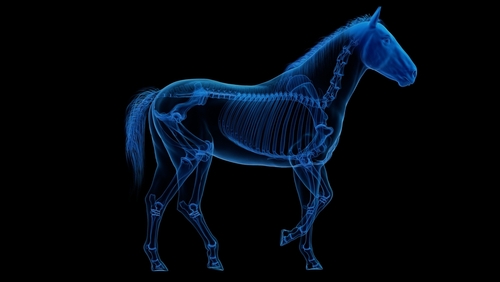
When your horse develops a shortened stride, persistent lameness, or an unexplained drop in performance, finding the cause starts with a thorough examination. At Chaparral Veterinary Medical Center in Cave Creek, we use equine diagnostic imaging to investigate problems beneath the surface and help guide treatment. Radiographs, ultrasound, and standing MRI each provide different information about your horse’s condition.
Radiographs, commonly called X-rays, help us evaluate bones and joints. We may recommend them to investigate fractures, arthritic changes, or other skeletal abnormalities. For horses with lameness, radiographs often provide an important starting point after we identify the area of concern.
Our imaging capabilities include digital radiography and specialized equipment for deeper structures, such as the spine and larger joints. We also have mobile digital radiography units for appropriate field evaluations. However, radiographs provide limited detail about many soft tissues, so additional imaging may be needed.
Ultrasound uses sound waves to create real-time images. It is especially useful for assessing tendons, ligaments, and other accessible soft tissues. We can examine tissue structure and look for changes that may help explain swelling or discomfort.
At our Cave Creek hospital, ultrasound also supports colic evaluations, internal medicine assessments, reproductive care, and cardiac imaging. The area being examined determines how we use it. Some structures, particularly those within the hoof capsule, can be difficult to assess fully with ultrasound.
Magnetic resonance imaging, or MRI, produces detailed images of both bone and soft tissue. It can help identify injuries within the hoof and other lower limb structures when radiographs or ultrasound have not fully explained the problem.
During a standing MRI, the horse remains standing under sedation, avoiding the need for general anesthesia for that scan. Keeping the horse sufficiently still is important for image quality. Our standing MRI capabilities include studies of the foot, fetlock, and suspensory ligament region, depending on the clinical concern.
We may recommend MRI after a lameness examination and diagnostic nerve blocks help localize the source of pain. It can reveal tendon or ligament damage and changes within bone that are difficult to detect with other tests.
We select imaging based on your horse’s history, examination findings, and the tissues we need to evaluate. Our approach may include:
Radiographs to investigate suspected bone or joint changes.
Ultrasound to assess accessible tendons, ligaments, and internal structures.
Standing MRI to investigate selected lower limb problems that need further clarification.
More than one method may be appropriate. We interpret imaging alongside the clinical examination, then use the findings to discuss treatment, rehabilitation, or further evaluation. For horse owners in Cave Creek, this helps connect the diagnostic process to practical decisions about care.
If your horse is showing signs of lameness or reduced performance, contact Chaparral Veterinary Medical Center in Cave Creek, AZ, by calling (480) 595-8600 to schedule an evaluation and discuss the right diagnostic imaging options.


…