Cave Creek, AZ 85331
span widget
span widget
Lameness in horses can be complex and frustrating for both owners and veterinarians. Whether it's a subtle change in gait or a pronounced limp, pinpointing the root cause is essential for effective treatment and recovery. Among the many diagnostic tools available, Magnetic Resonance Imaging (MRI) stands out for its precision and detail. But when is MRI the right choice for your horse?
Lameness refers to any abnormality in a horse’s gait caused by pain or mechanical dysfunction. Causes can range from soft tissue injuries and joint problems to bone fractures and hoof conditions. Traditional diagnostics such as physical exams, flexion tests, nerve blocks, and radiographs are often the first steps. However, when these methods do not provide a clear diagnosis, advanced imaging becomes necessary.
MRI provides detailed images of both soft tissue and bone, which sets it apart from other imaging tools like X-rays or ultrasound. While X-rays are best for visualizing bone and ultrasound is ideal for certain soft tissue structures, MRI combines both capabilities, making it especially useful in complex cases.
With MRI, veterinarians can identify:
Deep soft tissue injuries not visible on ultrasound
Early-stage bone bruising or stress fractures
Tendon and ligament injuries within the hoof capsule
Joint inflammation or subtle abnormalities
This level of detail allows for a more accurate diagnosis and a more targeted treatment plan.
MRI is typically considered when:
Lameness persists despite standard treatments
Nerve blocks have localized the issue but imaging has been inconclusive
The injury is suspected to involve deep structures within the hoof or lower limb
A precise diagnosis is needed to inform surgery or advanced therapies
At Chaparral Veterinary Medical Center, our team uses MRI to evaluate cases where conventional diagnostics fall short. It is especially valuable in diagnosing navicular syndrome, deep digital flexor tendon injuries, and collateral ligament damage.
Equine MRI can be performed on a standing sedated horse or under general anesthesia, depending on the equipment and the region being scanned. The process is safe and noninvasive, and our experienced veterinary team ensures your horse’s comfort throughout the procedure. Results are interpreted to guide the next steps in care.
While not every case of lameness requires an MRI, it can be a critical tool when other diagnostics fail to provide answers. Choosing MRI can lead to faster recovery, more effective treatment, and peace of mind.
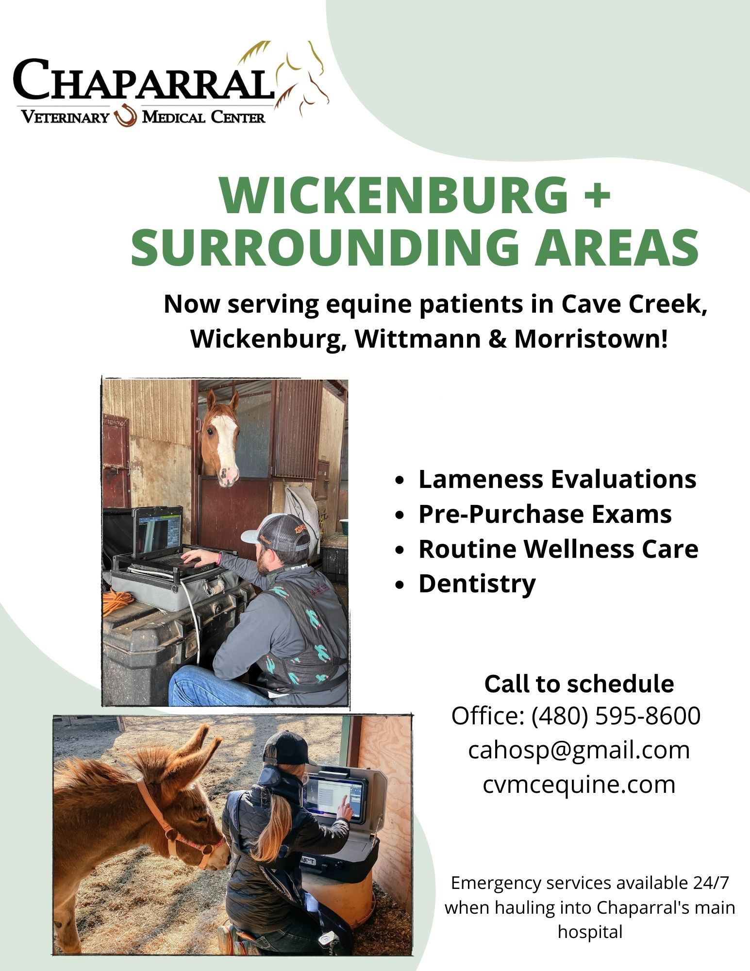
If your horse is experiencing unresolved lameness, schedule an evaluation with Chaparral Veterinary Medical Center to see if MRI is the right next step. Visit our facility in Cave Creek, Arizona, or call (480) 595-8600 to book an appointment today.


One fine body…